

QUICK LINKS: Practice Support Tools | Patients | Find a Neuro-Ophthalmologist | NOVEL | YONO Portal | Our Journal | Fellowships
Migraine Visual Aura
Patients: Download as PDF
Clinicians: Download as PDF
What is a migraine visual aura?
Migraine visual aura is the most common type of temporary neurological symptom and usually occurs just before or with the onset of a headache. It typically lasts about 30 minutes, but can range between 5 and 60 minutes. Migraines affect about 12% of people and among those with migraines, visual aura occurs in 25-30%. The frequency of the episodes may vary from person to person. The migraine visual aura can even occur without associated headache, which is more common over age 50.
A typical migraine episode has four parts: the premonitory phase (prodrome), the aura phase, the headache phase, and the post-headache phase (postdrome). Some of the phases may not be present in all people or in every migraine episode.
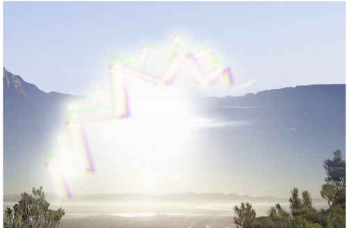 |
The visual pattern experienced may be different from one individual to another, but a common description is a fortification pattern (zigzag lines), scintillating or shimmering, with a crescent shape (“c” shape), often starting smaller in the center of the vision and then progressing towards the periphery (edge). It may simultaneously expand in size before eventually fading away. Variations include a progression from the periphery (edge) to the center, no progression at all, the presence of dots, lines or a missing spot in the visual field, and tunnel vision. The visual symptoms should be seen in both eyes, but symptoms tend to be more noticeable in the eye which has outer (lateral) visual field involvement.
After the episode, vision should return to the way it was before the episode. If it does not, one should seek medical evaluation. Some associated signs that suggest the need for an evaluation include a visual aura that always occurs on one side only, weakness or clumsiness during or after an episode, or headache occurring before the visual aura.
How can a neuro-ophthalmologist help?
Neuro-ophthalmologists are experienced and trained in assessing the characteristics of your visual symptoms and determining if they are usual or unusual for migraine visual aura as there are other vision disorders that can mimic migraine aura. The neuro-ophthalmological examination may also include formal visual field testing (peripheral vision testing) that can help distinguish migraine auras from other causes of visual disturbance. At the end of the evaluation, the neuro-ophthalmologist will be able to determine if additional investigations such as an MRI scan of the brain or other tests are needed.
Is there treatment for migraine visual aura?
The primary goal of migraine treatment is to address the headache. The visual aura is often resolved by the time the headache occurs, does not cause any permanent vision damage and is typically not the target of treatment. The treatment options vary from over-the-counter analgesics (pain medication) to migraine-specific prescribed medications such as the triptan class of medications. People who consistently have migraine visual aura followed by headaches can use the visual aura as an indicator that they should take their migraine medication quickly to stop the headache. When the migraine episodes are frequent, daily prophylactic medications can be used to decrease the frequency and prevent the episodes from happening. Although these prophylactic medications are not directed at the migraine visual aura, better control of the migraine attacks can also reduce the number of times the visual aura happens. When patients only experience visual aura and do not have headaches, typically no medication is recommended, although some patients find benefit from supplementation with vitamin B2 (riboflavin) and magnesium. You can consider discussing such treatment options with your doctor. A migraine journal can be kept in which you keep track of potential triggers, such as what you ate or drank, before migraine auras occur. After many months, you might be able to identify a common trigger that can be eliminated in order to reduce the frequency of the auras.
Additional Reading/Resources
- American Headache Society
https://americanheadachesociety.org/topic/migraine-with-aura/ - American Academy of Ophthalmology
https://www.aao.org/eye-health/diseases/what-is-migraine
Copyright © 2024. North American Neuro-Ophthalmology Society. All rights reserved.
This information was developed collaboratively by the Patient Information Committee of the North American Neuro-Ophthalmology Society. This has been written by neuro-ophthalmologists and has been edited, updated, and peer-reviewed by multiple neuro-ophthalmologists. The views expressed in this brochure are of the contributors and not their employers or other organizations. Please note we have made every effort to ensure the content of this is correct at time of publication, but remember that information about the condition and drugs may change. Major revisions are performed on a periodic basis.
This information is produced and made available “as is” without warranty and for informational and educational purposes only and do not constitute, and should not be used as a substitute for, medical advice, diagnosis, or treatment. Patients and other members of the general public should always seek the advice of a physician or other qualified healthcare professional regarding personal health or medical conditions.


.png)

_250x90.png)